|
|
 |
 |
Dr. Ashay H. Nandeshwar
M. D., (Ayu)(Mumbai)
(Podar Govt. Medical College)
P.G.D.M.L.T., L.Lb. (Sch)
Lecturer V.P.A.M.C.
Sangli |
| Read More |
|
|
|
|
 |
| Procedure 1: Preparing the blood smears |
| |
Distilled water was used to clean all the slides. The slides were then kept open to dry
5 ml syringes were used to take out blood from the patients. In adult patients, the blood was taken out from a vein in the arm; and in young children the blood was taken from finger tips.
About 0.5 ml of blood was taken out from each patient.
Each blood sample was then added into a test-tube (cleaned and dried).
Separate droppers were used to transfer 1 drop of blood from each sample from the test-tubes to the slides.
The slides were then covered with Leishman stain, and were then allowed to dry for about 30 minutes each. |
|
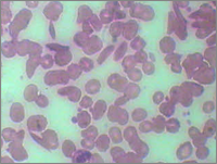
| Procedure 2: Viewing the smears under a microscope and counting the cells |
- The prepared slides were then viewed under an optical microscope.
- A digital camera was used to capture the images from the eye-piece.
- The digital images were then zoomed on a computer and then counted.
|
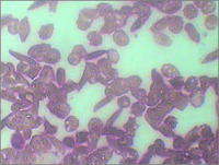
| At the start of treatment: |
|
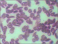
| Two months of treatment: |
|
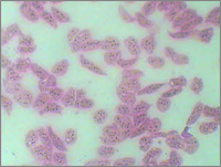
| 4 months of treatment: |
|
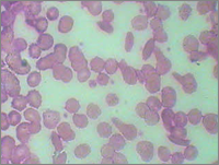
| 6 months of treatment: |
|
| 8 months of treatment: |
|
| |
|
|
|